EUS
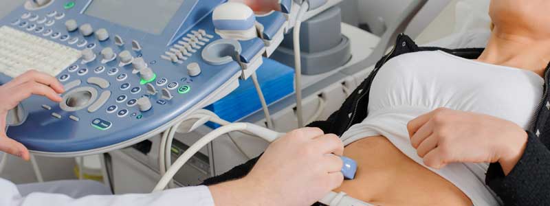
Endoscopic ultrasound is a procedure that combines endoscopy and ultrasound to create images of the digestive tract and nearby organs and tissues. It also is called EUS. During EUS, a thin, flexible tube called an endoscope is placed in the digestive tract. An ultrasound device on the tip of the tube uses high-frequency sound waves to create detailed images of the digestive tract and other organs and tissues. These include the lungs, pancreas, gall bladder, liver and lymph nodes. EUS helps find diseases in these organs and tissues and the digestive tract.
Sometimes a small needle is attached to the tube. This device is used to remove fluid or tissue that will be tested in a lab. This type of sample is called a biopsy. This procedure is called EUS-guided fine-needle aspiration or EUS-guided fine-needle biopsy. EUS also can be used to drain fluid from a lesion or deliver medicine to a specific place in the body.
EUS can capture images of other organs and nearby tissues too. They include:
Lungs.
Lymph nodes in the center of the chest.
Liver.
Gall bladder.
Bile ducts.
Pancreas.


